Scientist of the Day - Henry Vandyke Carter

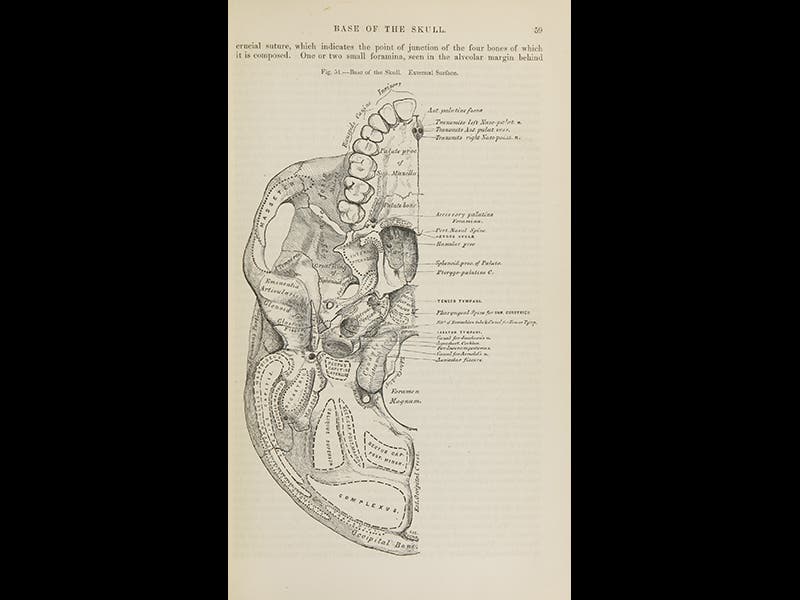
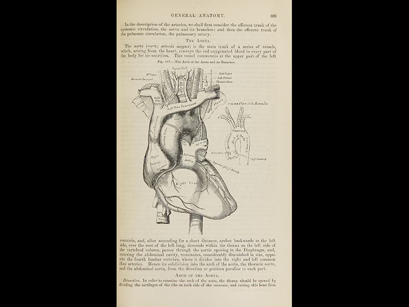
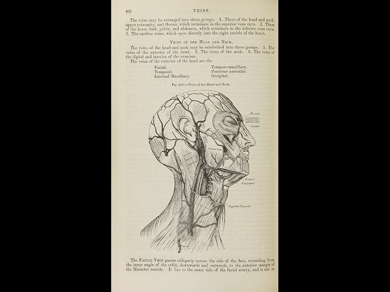
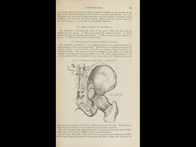
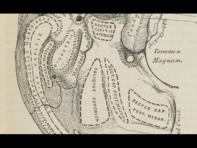
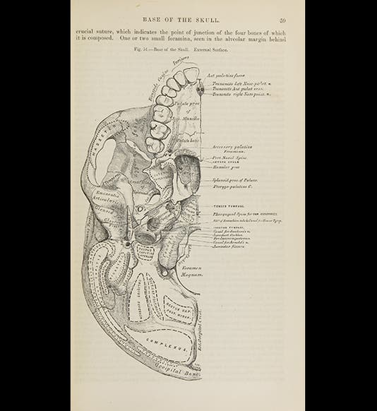
Henry Vandyke Carter, an English physician, surgeon, and artist, was born May 22, 1831. For two years, in 1856 and 1857, Carter labored night and day to produce 363 exquisite anatomical drawings which he drew directly on boxwood blocks ready for the wood engraver. The illustrations were intended to accompany a new anatomy text, written by his friend and fellow instructor at St. George's Hospital in London, Henry Gray. Because of a misunderstanding, most of the blocks were too big for their intended pages, and they had to be shaved, at great expense, and their engraved captions replaced with smaller type-set captions, which almost capsized the entire project. But the book finally appeared in September of 1858. The title on the title page was Anatomy, Descriptive and Surgical. The spine, however, said Gray's Anatomy, and Gray's Anatomy it has been ever since. The feature that endeared the book to students was the clarity of Carter's wood engravings, which were far superior to most anatomical illustrations before and since. But poor Carter got very little credit for his vital contribution, thanks mostly to Gray, who sent back the proofs of the title page and demanded that the font size for Carter’s name be reduced and his titles removed. In most modern editions of the book, Carter’s name doesn’t even appear. Perhaps the muse of justice arranged it so that Gray died in 1861, before he could enjoy much of his misappropriated fame, while Carter lived another 40 years and had quite a successful career. We do not have any edition of “Carter’s” Anatomy in the Library, ostensibly because human anatomy is out of scope for our collections, but since we have an exceptional collection of the history of scientific illustration, and since Carter played a major role in that history, one might think a case could be made for the acquisition of the first edition. The illustrations above are from the copy of the first U.S. edition (1859) at the National Library of Medicine and depict, in order: the base of the skull, the aorta, the veins of the skull, and the articulation of the pelvis. In addition, we include a detail of the base of the skull, so you can appreciate Carter’s ability to draw forth clarity from anatomical chaos. Dr. William B. Ashworth, Jr., Consultant for the History of Science, Linda Hall Library and Associate Professor, Department of History, University of Missouri-Kansas City. Comments or corrections are welcome; please direct to ashworthw@umkc.edu.