Scientist of the Day - Camillo Golgi
Camillo Golgi, an Italian physician and pathologist, was born July 7, 1843. Golgi worked as the resident physician at a mental hospital between Milan and Pavia in Italy, and he spent his spare time trying to unravel the mysteries of the nervous system, the complexity of which had stymied researchers prior to Golgi. By Golgi's time, the cell theory was well established, and physiologists understood that all body tissues and organs are comprised of cells, butt the nervous system seemed to be an exception. Many thought that the nerves formed an extended reticular network that could not be sub-divided into cells. The problem was difficult to solve, because individual nerve cells were all but invisible in the microscopes of the time, and nothing whatsoever could be resolved in complex nervous tissue such as the brain. Golgi spent years tinkering with stains and dyes that might make the nerves stand out against the surrounding tissue. Eventually, in 1873, he stumbled on the discovery that, if one dipped a slice of brain tissue in potassium dichromate (which hardened it) and then into a silver solution (silver nitrate), the nerve cells were stained black (Golgi called it "the black reaction"). Better yet, only a few random nerve cells accepted the stain, so that they stood out starkly from the surrounding cells. For the first time, one could see an actual nerve cell, with its lengthy axon, and all the dendrites. We include here one of Golgi’s drawings of neurons darkened with silver (second image, below). This staining technique is now called the Golgi method, and it has been invaluable in studying the nervous system ever since. Interestingly, Golgi concluded that the reticular theory is correct, and that the nerves form a continuous network that runs unbroken throughout the body.

One of Camillo Golgi’s drawings of neurons darkened with silver (nlm.nih.gov)
Twenty years after Golgi published his method, a Spaniard, Santiago Ramón y Cajal, used it to show that Golgi was wrong about the reticular theory. The nervous system is not continuous but is formed of individual nerve cells with their long axons and numerous dendrites, and these cells are interrupted by synapses, across which the cells must communicate. This came to be called the neuron doctrine. The nervous system was no longer an exception to the cell theory; everything in a living organism is indeed comprised of cells. In 1906, Golgi and Ramón y Cajal were jointly awarded the Nobel Prize in Physiology/Medicine for founding the science of neurology and discovering the cellular nature of the nervous system. Golgi's reaction to receiving a prize for a theory he resisted all his life is not recorded.

Modern textbook illustration of a eukaryote cell and its organelles, with the Golgi apparatus clearly labelled (Ladyofhats on Wikimedia commons)
But Golgi had one more arrow in his quiver. In 1897, using the same staining techniques he had used on nerve cells, he discovered a little stack of membranes that is present in all animal and plant cells. You probably had to memorize its name for high-school biology class: it is called the Golgi apparatus, and it shares its cellular space with ribosomes, mitochondria, and the like. The Golgi apparatus (also called the Golgi body or Golgi complex) is responsible for packaging newly minted proteins for delivery outside the cell. In a diagram of a cell interior (third image, above), you can see the Golgi body in green at bottom left. It is odd that this organelle was discovered by Golgi, since he was not a cell biologist but a pathologist and really had no interest in the internal structure of the cell.

Sala Golgi, room devoted to memorabilia of Camillo Golgi, University of Pavia History Museum, Pavia, Italy (Museo per la Storia dell'Università, Pavia)
The University of Pavia is proud of their graduate and is undismayed that Golgi was unable to use his revolutionary technique to discover the neuron doctrine and that a Spaniard had to step in to help out. For a long time, in the University of Pavia History Museum, they had a large room (the Sala Golgi) devoted to Golgi memorabilia (fourth image, just above). There was a small statue of Golgi in the room, as well as the microscopes he used and many of his drawings and papers. I am not sure if the room is still intact, because the University has recently opened a separate Museo Golgi, and I do not know if the contents of the Sala Golgi were transferred to the Museo Golgi. But the images of the Sala Golgi are still up on the web, so I include two of them here.

Nobel Prize certificate awarded to Camillo Golgi in 1906, in the Sala Golgi, University of Pavia History Museum (Museo per la Storia dell'Università, Pavia)
The room also holds, or held, the actual Nobel certificate for Golgi’s Nobel Prize in Physiology/Medicine of 1906. Nobel medals are often seen, but it is rare to find an awarded certificate on display (fifth image, above).

Statue of Camillo Golgi at the University of Pavia (Museo per la Storia dell'Università, Pavia)
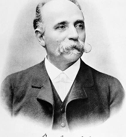
There is a larger statue of Golgi outside in a university courtyard (sixth image, above). But I think Golgi looks most distinguished in the formal photograph, taken before 1903, that we use as our first image. It is not surprising that this photograph was the one used on the Nobel prize webpage for the 1906 award.
Dr. William B. Ashworth, Jr., Consultant for the History of Science, Linda Hall Library and Associate Professor emeritus, Department of History, University of Missouri-Kansas City. Comments or corrections are welcome; please direct to ashworthw@umkc.edu.