Scientist of the Day - Volcher Coiter

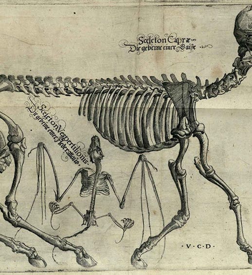
Skeletons of a goat and a bat, detail of a larger engraved plate, drawn by Volcher Coiter, in his Lectiones Gabrielis Fallopii de partibus similaribus humani corporis, plate 3, 1575, unknown copy (Wikimedia commons)
Volcher Coiter, a Dutch physician and anatomist from Groningen, died June 2, 1576, at the age of about 42. He learned anatomy at Padua, studying with two anatomists of tubular renown, Gabriele Falloppio and Bartolomeo Eustachi, and he studied zoology with Ulisse Aldrovandi in Bologna. With that background, it is not surprising that Coiter came to believe that anatomy applied to zoology might be a useful discipline. The tradition among natural historians was to omit discussion of internal anatomy when describing animals, and you will be hard pressed to find any images of skeletons or internal parts in the works of Conrad Gessner or Guillaume Rondelet, published in the 1550s. It was Coiter who first suggested that students of nature should study animals from the inside out, and he prepared a set of four large anatomical plates that he published in 1575. These plates, which he drew himself, depict the skeletons of such diverse animals as a tortoise, a goat, a weasel, and several birds, including a crane, a cormorant, and a starling. These engravings are astonishingly good, when you consider that they had no real precedent in zoology.

Portrait of Volcher Coiter, unknown artist, unknown date, German National Museum, Nuremberg (Wikimedia commons)
Colcher's works, of which there are only two, are quite scarce. The first, Externarum et internarum principalium humani corporis partium tabulae (1572), is concerned with human anatomy, with the only plates being, with one exception, copies or adaptations of woodcuts from Andreas Vesalius. His Lectiones Gabrielis Fallopii de partibus similaribus humani corporis was published in 1575. These contain the anatomy lectures of his teacher Falloppio, and the title continues: … his accessere diversorum animalium sceletorum explicationes iconibus artificiosis, et genuinis illustratae (... to which are added Explanations of Different Animal Skeletons, Illustrated with Contrived and Genuine Images). It is this latter work that contains the four large fold-out engravings with which we illustrate this post.

Skeletons of a crane, a cormorant, a starling, and a lizard, engraving by Volcher Coiter, in his Lectiones Gabrielis Fallopii de partibus similaribus humani corporis, plate 4, 1575, unknown copy (Wikimedia commons)
I first saw Coiter's four engravings when I was at the University of Wisconsin as a graduate student, and I was blown away by their high quality. I took slides of the four plates, which I still have, now in digital form, but they are, alas, of graduate student quality. I have never seen another copy. I was prepared to use my slides to illustrate this post, but I found that someone has uploaded all four plates to Wikimedia commons. As happens all too often, the uploader neglected to provide us with the location of the copy that was scanned. But they are high-resolution images of excellent quality, much better than my own photographs, and I see no problem in using them here, even if I cannot give them a proper source attribution.

Skeletons of a fox, a squirrel, hedgehog, frog, mouse, and mole, engraving by Volcher Coiter, in his Lectiones Gabrielis Fallopii de partibus similaribus humani corporis, plate 2, 1575, unknown copy (Wikimedia commons)
Detail of the skeleton of a mole (talpa), also showing its pectus, or powerful shoulder girdle, detail of fourth image, in Lectiones Gabrielis Fallopii de partibus similaribus humani corporis, by Volcher Coiter, plate 2, 1575, unknown copy (Wikimedia commons)
One of the plates is shown only in detail (first image), but we reproduce the other three in their entirety. In addition, we provide a detail of the second plate (our fourth image), where we can see the skeleton of a mole (talpa), as well as an added view of its shoulder girdle, which powers its digging digits and which apparently intrigued Coiter. Note that each plate is signed: “V.C.D.” – “delineated by Volcher Coiter.”

Skeletons of a piglet, a marten, a parrot, and a rabbit, engraving by Volcher Coiter, in his Lectiones Gabrielis Fallopii de partibus similaribus humani corporis, plate 1, 1575, unknown copy (Wikimedia commons)
Coiter was a contemporary of Ulisse Aldrovandi. But Coiter died young and Aldrovandi published late. We can see the impact of young Coiter on the later Aldrovandi if we examine Aldrovandi’s volumes on mammals and birds, some published after his death in 1605, where we occasionally see skeletons of animals accompanying the more traditional woodcuts of animal exteriors. We show you a woodcut from Aldrovandi’s De quadrupedibus digitatis (On Four-footed Animals with Claws, 1645), depicting a hedgehog skeleton (seventh image). If we look at a detail of Coiter’s second plate (eighth image), we see exactly where Aldrovandi found his illustration.

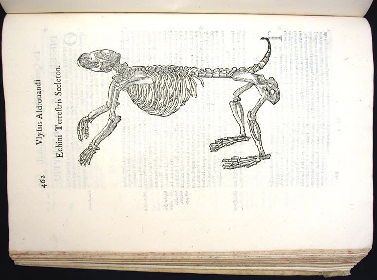
Skeleton of a hedgehog (echinus), woodcut, in Ulisse Aldrovandi, De quadripedibus digitatis viviparis, 1645 (1663) (Linda Hall Library)
The skeleton of a hedgehog (echinus), detail of fourth image, in Lectiones Gabrielis Fallopii de partibus similaribus humani corporis, by Volcher Coiter, plate 2, 1575, unknown copy (Wikimedia commons)
The portrait of Coiter, which we could only find in a second-hand copy (second image) is in the German National Museum in Nuremberg, where Coiter was city physician after 1569 and where his books were published.
William B. Ashworth, Jr., Consultant for the History of Science, Linda Hall Library and Associate Professor emeritus, Department of History, University of Missouri-Kansas City. Comments or corrections are welcome; please direct to ashworthw@umkc.edu.