Scientist of the Day - William Cheselden


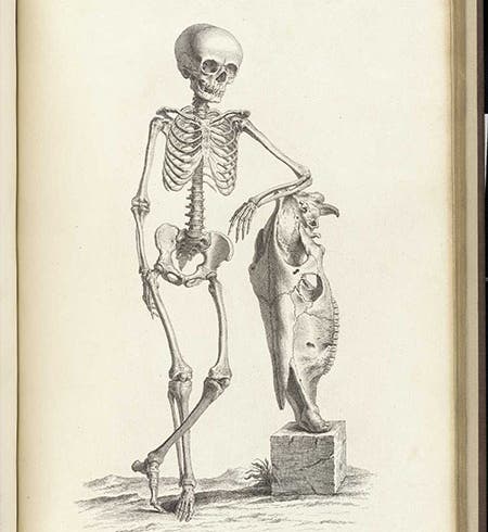
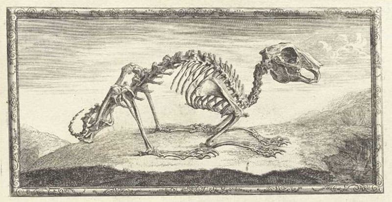
Skeleton of a 9-year-old boy with the skull of a horse, engraving, plate 33, in Osteographia, or the Anatomy of the Bones, by William Cheselden, 1733, National Library of Medicine (nlm.nih.gov)
William Cheselden, an English surgeon, died Apr. 10, 1752, at age 63. Cheselden was quite a famous surgeon in his day, noted for his expertness (and speed) in excising bladder stones, and for his skill in eye surgery and cataract removal. He apparently wore the turban shown in his portrait while at the operating table, and, like some thespians, was physically sick before each operation. Cheselden was openly critical of the control over surgery exerted by the medieval Company of Barbers and Surgeons, and he engineered a breakaway in 1745, which separated the two groups, producing the Company of Surgeons, which later (1800) became the Royal College of Surgeons.

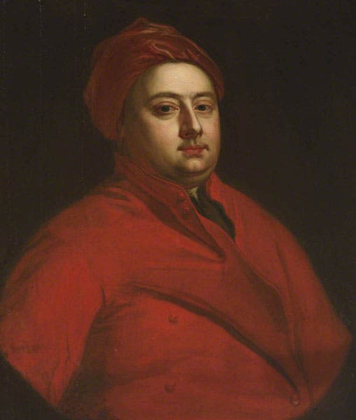
Portrait of William Cheselden, oil on canvas by Jonathan Richardson the Elder, undated, St. Thomas Hospital, London (artuk.org)
Outside the surgical community, Cheselden is best known for an illustrated book of anatomy that he published in 1733, called Osteographia, or the Anatomy of the Bones. He had earlier published an anatomy textbook that was widely used, but there the word took precedence over the image. He now had in mind a book where the image would be king, and where every bone in the human body would be shown life size, requiring very large plates, some 21 inches high. The book was expensive to produce, which necessitated a high price, resulting in low sales, so that less than 100 of the 300 copies printed were sold. We do not have a copy of the 1733 edition in our collections – it is out of scope for us – but we do have a full-size facsimile that was published in 1968, which we keep on the large-folio shelves in the rare book room. However, our illustrations today are taken from the copy in the National Library of Medicine, which is available online.

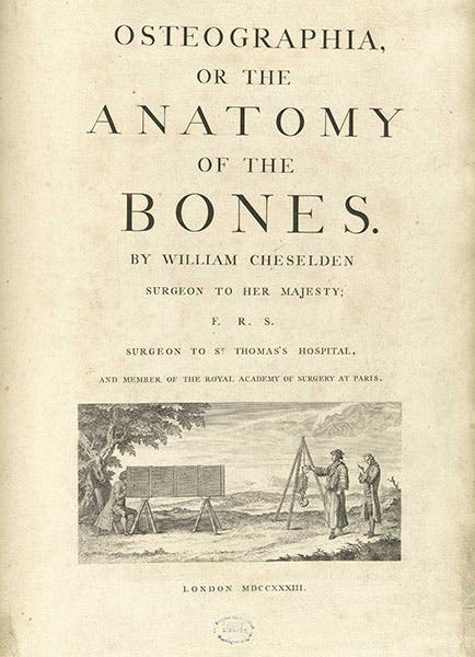
Title page with engraved vignette, Osteographia, or the Anatomy of the Bones, by William Cheselden, 1733, National Library of Medicine (nlm.nih.gov)

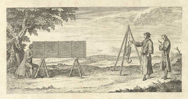
Title-page vignette, showing the use of a camera obscura in drawing the bones, engraving in Osteographia, or the Anatomy of the Bones, by William Cheselden, 1733, National Library of Medicine (nlm.nih.gov)
Leafing through the book is indeed a visual, if not visceral, experience, as there are 56 plates of human bones, with labels, and then a repeat of all 56, this time untainted by lettering. The preceding text is divided up into 8 short chapters, and for each of these there is an engraved frontispiece, an illustrated headpiece, and a decorative initial letter – all designed and executed especially for this book. The title page (third image) bears a vignette that depicts a large camera obscura, which Cheselden required that his artists use, projecting the bones and skeletons onto a ground glass drawing surface, from which the images were traced onto paper (fourth image).

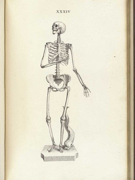
Female skeleton, in the pose of the Venus pudica (modest Venus), engraving in Osteographia, or the Anatomy of the Bones, by William Cheselden, plat 34, 1733, National Library of Medicine (nlm.nih.gov)
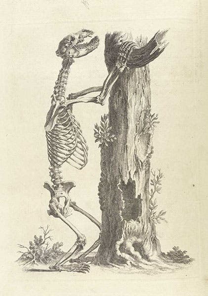
There are plates that show the bones of the skull, the jaw, the hands and feet, which we pass over here, since we can't show everything, but which you can view online. The complete skeletons had to be reduced in size, except for the fetal skeleton. There was a tradition in anatomical illustration, given the firm stamp of approval by Andreas Vesalius in his De fabrica (1543), that human skeletons should be set in landscapes, and Cheselden followed this to some extent, but not always. Few of his skeletons evoke the poses of Vesalius (to which he pays homage in the headpiece to the introduction, sixth image), but Cheselden was partial to Vesalian whimsy, as with the nine-year-old boy leaning on a horse skull that is half his size (first image), or the female skeleton in the pose of the Venus pudica, the modest Venus, where Cheselden preserved the hand that covered the now-missing breasts, but moved the other hand of modesty to the side, so that the pelvic bones would be visible (fifth image).

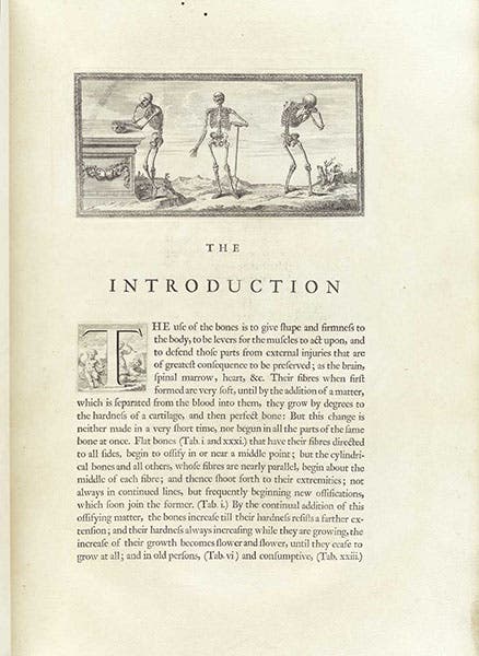
First page of the Introduction, with engraved headpiece, Osteographia, or the Anatomy of the Bones, by William Cheselden, 1733, National Library of Medicine (nlm.nih.gov)

Bear skeleton, frontispiece to chapter 4, engraving in Osteographia, or the Anatomy of the Bones, by William Cheselden, 1733, National Library of Medicine (nlm.nih.gov)
One surprise is that Cheselden stressed the importance, for the student of human anatomy, of comparative anatomy – the anatomy of animals. He did this by using animal skeletons for the frontispiece to each section, as well as for many of the decorative headpieces. There is an upright bear skeleton before chapter four (On the bones of the upper arm, seventh image), while a rabbit presides as the headpiece on the page opposite (eighth image), and a chameleon skeleton serves as the headpiece introducing chapter six (Cartilages, ligaments, &c, ninth image).

The skeleton of a rabbit, engraved headpiece to chapter 4, Osteographia, or the Anatomy of the Bones, by William Cheselden, 1733, National Library of Medicine (nlm.nih.gov)

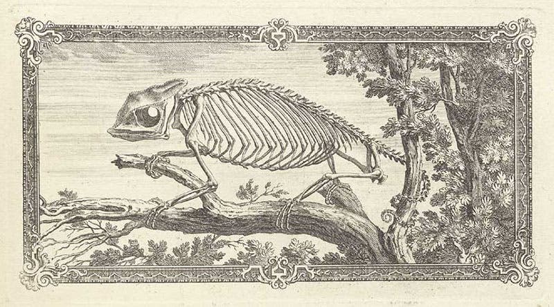
The skeleton of a chameleon, engraved headpiece for chapter 6, Osteographia, or the Anatomy of the Bones, by William Cheselden, 1733, National Library of Medicine (nlm.nih.gov)
The initial letters to the eight chapters are hard to see at this scale, but they follow another initiative of Vesalius, who used specially cut woodcut initials that showed putti engaged in anatomical exercises, such as boiling bones or dissecting animals. Cheselden’s engraved initials are smaller and thus less detailed, but they are charming nonetheless. I tried to have one enlarged sufficiently for you to view, the initial for the first page of the introduction (sixth image), where several putti steal a skeleton from a grave in a cemetery (tenth image).


An engraved initial letter “T”, showing putti digging up a skeleton in a graveyard, detail of the first page of the Introduction to Osteographia, or the Anatomy of the Bones, by William Cheselden, 1733, National Library of Medicine (nlm.nih.gov)
I like Cheselden’s Osteographia because it does not immediately call to mind any earlier book of anatomy, the way most 17th-century anatomy books recall the work of Vesalius. Govard Bidloo’s anatomical book of 1685 broke radically with the past, and Cheselden’s, while paying respects to the Vesalian tradition, did the same. That is not easy to do and do well. The Osteographia deserved wider sales and better renown than it received at the time, but which it is getting now. There was an excellent article on Cheselden and his Osteographia in the Public Domain Review, an online journal that caters to studies of image-driven science. The article, by Monique Kornell, was published in 2011; you may access it here.
William B. Ashworth, Jr., Consultant for the History of Science, Linda Hall Library and Associate Professor emeritus, Department of History, University of Missouri-Kansas City. Comments or corrections are welcome; please direct to ashworthw@umkc.edu.